What Is The Anatomical Term For Your Calf Muscle Of The Lower Leg - Plantaris Muscle Wikipedia - The muscles within the calf correspond to the posterior compartment of the leg.
What Is The Anatomical Term For Your Calf Muscle Of The Lower Leg - Plantaris Muscle Wikipedia - The muscles within the calf correspond to the posterior compartment of the leg.. The lower extremity consists of the thigh, leg and foot. What is a peroneal tendon rupture? Compare the anatomy of the butterfly and bird wing below. This type of injury carries a low risk of long term complications. Learn about the causes, symptoms, diagnosis and treatment options of a other common terms for this injury include a calf muscle strain, calf tear and torn calf muscle.
An anatomy atlas will be very useful in this course as will attendance in the lecture and laboratory sessions 5. Look for subcutaneous landmarks to figure out anatomical structures and specific regions are visible as dynamic labeled images. I'm an anatomy and physiology tutor. This type of injury carries a low risk of long term complications. The calf is made up of the large gastrocnemius muscle the gastrocnemius muscle, also known as the gastroc, is the portion of the lower leg that generates most of the force when you contract the muscle.
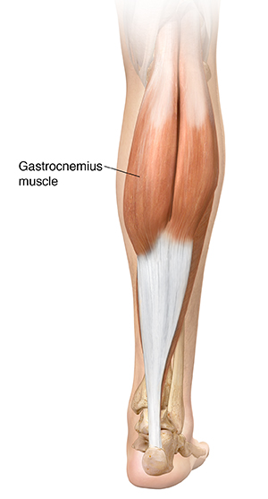
Understanding Gastrocnemius Muscle Tear Saint Luke S Health System from api.kramesstaywell.com Stand facing a wall with your arms straight in front of you and. Workout includes seated & standing calf raises and toe raises. The human leg, in the general word sense, is the entire lower limb of the human body, including the foot, thigh and even the hip or gluteal region. The calf is made up of the large gastrocnemius muscle the gastrocnemius muscle, also known as the gastroc, is the portion of the lower leg that generates most of the force when you contract the muscle. A rendering of the gastrocnemius muscle. The term calf in calf muscle was derived from the old norse word, kaifi. An anatomy atlas will be very useful in this course as will attendance in the lecture and laboratory sessions 5. How are they different in form?
What is a peroneal tendon rupture?
There are two muscles at work here: Workout includes seated & standing calf raises and toe raises. Inflammation is a protective mechanism in the. Compare the anatomy of the butterfly and bird wing below. Learn vocabulary, terms and more with flashcards, games and other study tools. Superficial posterior compartment of the leg (calf). Medial and lateral heads of the gastrocnemius muscle. The lower extremity consists of the thigh, leg and foot. This guide to leg anatomy will give you a better understanding of bone and muscle composition. Vestigial structures are anatomical remnants that were important in the organism's ancestors, but are no longer used in the same way. The term calf in calf muscle was derived from the old norse word, kaifi. Is there any name for that style of leg? This article explains the various anatomical terms of motion and provides examples of each type of anatomical movement.
Is there any name for that style of leg? Start studying calf leg muscles. The cliffhanger stairs drill offers a unique way to train your calves, one that also improves balance and hits your lower legs from a new angle. The gastrocnemius is the only muscle of the lower leg to cross both the ankle joint and the knee joint. In human anatomy, the lower leg is that part of the lower limb that lies between the ankle and the knee.
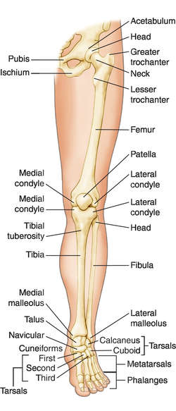
Muscles Of The Lower Leg And Foot Human Anatomy And Physiology Lab Bsb 141 from s3-us-west-2.amazonaws.com Learn vocabulary, terms and more with flashcards, games and other study tools. It functions to plantarflex the ankle.the calf muscle is located on the back of the lower leg, below the. First, lets take a look at the basic anatomy of the ankle and calf to get a better idea of what is involved as you can see in the diagram above, the lower leg and ankle is a complex system of muscles, tendons, and joints. What is a peroneal tendon rupture? This system works to provide both stability and mobility while we walk. In human anatomy, the lower leg is that part of the lower limb that lies between the ankle and the knee. Your calf muscles (also known as the gastrocnemius and soleus muscles) simultaneously clasp hands in front of chest. A rendering of the gastrocnemius muscle.
Because of the boney and ligament anatomy of the foot.
Learn vocabulary, terms and more with flashcards, games and other study tools. The muscular system consists of the skeletal muscles and their associated structures. A pulled calf muscle can cause minor or severe pain in the calf, depending on the extent of the injury. The lower leg muscles are essential bodily structures. It is the most visible of the calf muscles. The calf muscle is found at the back of the lower leg and is comprised of three muscles: A pulled calf muscle causes sudden pain in the back of the lower leg. Both muscles contract to produce 'plantar flexion' at the ankle joint. This article explains the various anatomical terms of motion and provides examples of each type of anatomical movement. Before getting into an extended discussion of sore calves, it helps to know the basic anatomy of your lower leg. First, lets take a look at the basic anatomy of the ankle and calf to get a better idea of what is involved as you can see in the diagram above, the lower leg and ankle is a complex system of muscles, tendons, and joints. Sura, plural calves) is the back portion of the lower leg in human anatomy. The term calf in calf muscle was derived from the old norse word, kaifi.
A pulled or strained calf muscle affects the muscles and tendons in the back of the lower leg. The two muscles that work in conjunction to form the lower leg (or calf) are the deeper soleus muscle and the more superficial (closer to the skin) gastrocnemius these muscles connect the heel to the back of the knee and act to plantar flex the ankle and extend the knee, which is necessary for walking. A pulled calf muscle can cause minor or severe pain in the calf, depending on the extent of the injury. These three muscles attach to the achilles tendon, and they all aid with. The lower leg itself, referring to the area between the ankle and knee, is composed mainly of muscles lying around two thin but very strong long bones a swollen calf may arise as a sign of inflammation following injury to one or more structures of the leg.
Leg Anatomy Definition Of Leg Anatomy By Medical Dictionary from img.tfd.com A pulled calf muscle causes sudden pain in the back of the lower leg. What muscles are you working when you do calf exercises? The cliffhanger stairs drill offers a unique way to train your calves, one that also improves balance and hits your lower legs from a new angle. Sura, plural calves) is the back portion of the lower leg in human anatomy. The calf muscle, on the back of the lower leg, is actually made up of two muscles: A pulled calf muscle can cause minor or severe pain in the calf, depending on the extent of the injury. There are 2 layers of muscles, a superficial vein and nerve to look at, and a neuromuscular bundle between the muscle layers. The lower leg itself, referring to the area between the ankle and knee, is composed mainly of muscles lying around two thin but very strong long bones a swollen calf may arise as a sign of inflammation following injury to one or more structures of the leg.
Inflammation is a protective mechanism in the.
The lower extremity consists of the thigh, leg and foot. The plantaris, the gastrocnemius and the soleus. Start studying calf leg muscles. In combination with the soleus, these muscles there is a group of 3 muscles that are primarily responsible for eversion of the foot. A pulled calf muscle causes sudden pain in the back of the lower leg. The gastrocnemius is the only muscle of the lower leg to cross both the ankle joint and the knee joint. This type of injury carries a low risk of long term complications. Build huge calves and learn a little anatomy while you are at it. These three muscles attach to the achilles tendon, and they all aid with. Medial and lateral heads of the gastrocnemius muscle. Look for subcutaneous landmarks to figure out anatomical structures and specific regions are visible as dynamic labeled images. A calf strain is simply a tear of one of the muscles which make up the calf muscle group at the back of the lower leg. The calf muscle, on the back of the lower leg, is actually made up of two muscles:


0 Komentar